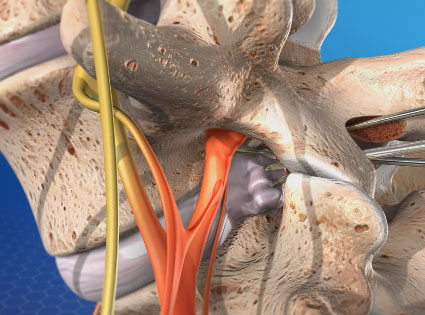
Lumbar disc microsurgary is a minimally invasive surgery to correct problems in the lower back such as a slipped, or herniated disc. A slipped disc can lead to pain when it presses on a nerve in the lower back, especially the long sciatic nerve. This nerve is the longest in the body and bifurcates to travel down the lower part of the spinal column, down the buttocks and down the legs to the feet. The intense, shooting pain and accompanying numbness and tingling can interfere with the quality of a patient’s life.
Human beings often have trouble with their lower, or lumbar back because of the load this part of the back has to bear.
The surgery is called microsurgery because it doesn’t rely on the open surgery that is traditionally used to treat herniated or slipped discs. Open surgery requires a long incision in your back. Muscles and other tissue need be pushed aside or even cut in order to access your spinal column.
During lumbar disc microsurgary the surgeon makes tiny incisions and uses miniaturized surgical tools to operate. They also make use of a special microscope. This reduces the trauma to the muscles and tissues around the spinal column and allows the doctor to only remove the part of the herniated disc that is impinging on your spinal nerve and causing your pain. The rest of the disc can be left intact.
Anatomy of the Vertebra
Before describing lumbar disc microsurgery, it is useful to describe the vertebra. Though a baby is born with 33 vertebrae, they fuse over the years until an adult has 26. The vertebra has a central body with a pedicle or extension on each side. The pedicles join bilateral laminae to make an arch. The arch encloses an opening called the foramen, and the spinal cord passes through the foramen. The spinous process and two transverse processes extend from the arch. The transverse processes have articular processes that connect to the articular processes of other vertebra.
The Surgery
During lumbar disc microsurgary, the surgeon makes a tiny incision in the lamina right over the slipped disc. The surgeon uses a microscope to find the compressed nerve and the disc. Then, they move the spinal nerve away from the disc with a nerve retractor. This frees up space for them to work. The surgeon only removes the part of the disc that’s impinging on your spinal nerve. In an open surgery, the entire disc is removed and adjoining vertebrae are encouraged to fuse through the insertion of cages or plates.
After microsurgery, the surgeon replaces the nerve, sutures the surgical wound and covers it with dressings. Because the surgery was minimally invasive, your recovery time is much shorter than would be if you’d had the open type of lower back surgery. You may not even need to spend the night in the hospital but can go home after a period of rest.
You can recuperate at home, and go back to work after about two weeks if your work isn’t too strenuous. You can return to the gym after about a month or a month and a half.
Call Dr. Courtney at Advanced Spine Center in Plano today!
At the Advanced Spine Center, we know that the single most important person in the process of spine care is you, the patient. Well-informed patients are more successful in following through on their treatment and have better outcomes. The first step to success is in knowing what to expect. We invite you to contact us today and schedule a consultation with Dr. Courtney to discuss any questions you may have regarding cervical disc disease or any other spine condition.
When it comes to spine care in Plano, you want and deserve the best-of-the-best!

